People DO Write Songs about our group's research:
"I Study Chromium" by former undergrad Ernesto Abruna:
Research Directions:
Orbital Domain Dynamics Below theVerwey Transition
Mesoscale phenomena play an important role in the dynamics of phase transitions in strongly
correlated electron systems. In addition to the coupling of charge, spin, orbital, and
lattice degrees of freedom, the underlying mechanism of phase transformations is influenced
by nanoscale heterogeneities such as local strain fields, phase separation, domain walls,
and defects. Magnetite (Fe3O4) is a model correlated electron system
that exhibits a metal-insulator transition at T = 123 K that is
simultaneously accompanied by a structural phase transition. Despite intense research since
its discovery by Verwey in 1939, the role that electronic dynamics and mesoscale phenomena
play in driving the transition is poorly understood.
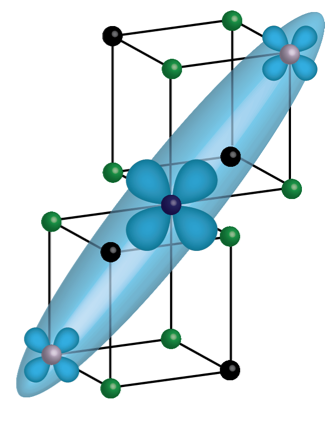
Charge and orbital ordering associated with the trimeron, three-site lattice
distortions, where Fe+2 (blue) sits at the center of the
trimeron. Fe3+ (gray) atoms sit at the apex of the trimeron, and
oxygen atoms are in green.
We used X-Ray Photon Correlation Spectroscopy (XPCS) to directly probe the
electronic structure of magnetite. By tuning to the Fe L3 edge, we are able to
access a forbidden (001/2) superlattice peak that is a direct signal from the
t2g orbitally ordered domains present in the low temperature insulating
phase. The speckles are fingerprints of the orbital domains, and by observing how
fast the speckles change over time, we gain valuable information to the role orbital
structure plays in driving the metal-insulator transition. Surprisingly, the fastest
dynamics occur at T = 90 K, well below the transition temperature.
Combined with the peak intensity and peak width data, our measurements reveal the
dynamics below T = 90 K can be ascribed to an Arrhenius behavior,
indicating a thermally driven effect, whereas the dynamics above
T = 90 K is dominated by the onset of phase separation of metallic
and insulating domains.
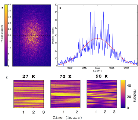
(a) Detector image of the speckle pattern from magnetite. (b) Gaussian fit across the speckle pattern to obtain the size of the orbitally ordered domains. (c) Waterfall plots that visually demonstrate how the individual speckles evolve over time.
Reference:
Roopali Kukreja, Nelson Hua, Joshua Ruby,
Andi Barbour, Wen Hu, Claudio Mazzoli, Stuart Wilkins, Eric E. Fullerton, and Oleg
G. Shpyrko, "Orbital Domain Dynamics in Magnetite below the Verwey
Transition,"
Phys. Rev. Lett.
121, (2018).
|
Nucleation of dislocations and their dynamics in layered oxide cathode materials
during battery charging
Lithium-rich layered oxides (LRLO) are among the leading candidates for the
next-generation cathode material for energy storage, delivering 50% excess
capacity over commercially used compounds. Despite excellent prospects, voltage
fade has prevented effective use of the excess capacity, and a major challenge
has been a lack of understanding of the mechanisms underpinning the voltage
fade. Using operando three-dimensional Bragg coherent diffractive imaging we
directly observe the nucleation of a mobile dislocation network in LRLO
nanoparticles. The dislocations form more readily in LRLO as compared with a
classical layered oxide, suggesting that the formation of partial dislocations
contributes to the voltage fade.
|
|
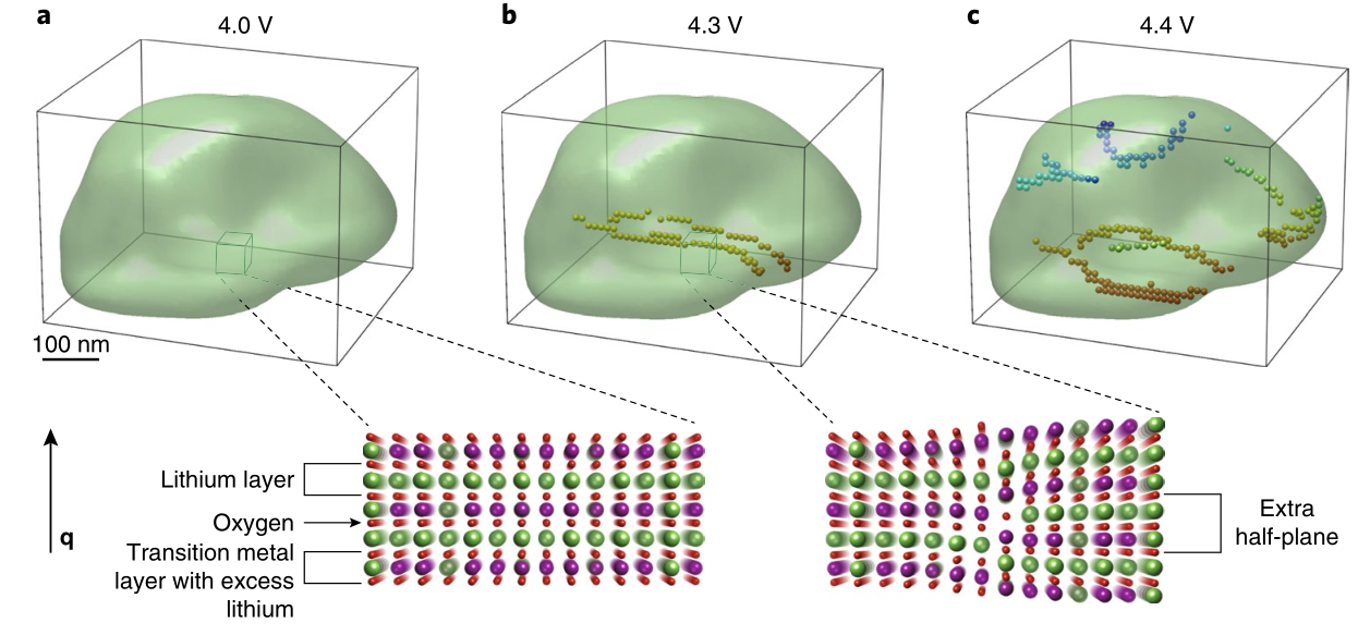
Formation of a dislocation network during charge. Isosurface rendering
of a LRLO particle measured in operando during charge. (a) At a charge
state of 4.0 V vs. Li+ no dislocations are observed in
the particle. The inset shows a schematic of a dislocation-free
crystal.(b) At 4.3 V two edge dislocations have formed during
lithium extraction (shown by small spheres in the particle). The inset
shows a schematic of an edge dislocation. (c) At 4.4 V a
dislocation network emerges (colours are used to represent different
dislocations). The direction of the X-ray scattering vector q
(perpendicular to the layers) is indicated and the size of the particle
is around 300×300×500 nm3.
|
|
The experimental observation of a significantly higher rate of dislocation
formation in LRLO as compared with classical material resonates with the excess
capacity in LRLO. As our X-ray data show no evidence of a two-phase composite,
we rule out excess capacity due to the activation of monoclinic
Li2MnO3 at high voltages. The current understanding of the
anionic activity in LRLO materials includes three processes: step 1 — reversible
oxidation of O2− to O− ; step 2 — further partially
reversible oxidation of oxygen; and step 3 — irreversible release of
O2 gas from the bulk material to the surface. Steps 2 and 3 require
elevated oxygen mobility in the bulk. While enhanced ‘pipe’ diffusion occurs
along dislocations, recent theoretical works suggest a slower transport of
oxygen along dislocations due to cation charge accumulation at the defect site,
with oxygen mobility due to thermal fluctuations predicted to be negligible at
room temperature. However, an external electrical current leads to reversible
accumulation or depletion of oxygen vacancies at dislocations in SrTiO
3. Dislocations play a major role in redox-based resistive switching
in transition metal oxides used for memristive applications. Similarly, we posit
that during charge the lithium and electron extraction activates the emergent
dislocation network in LRLO for the transport of oxygen vacancies. The oxygen
vacancy mobility is probably accommodated by undesired side effects such as
cation disorder and stacking faults, which debilitate oxygen redox activity.
|
|
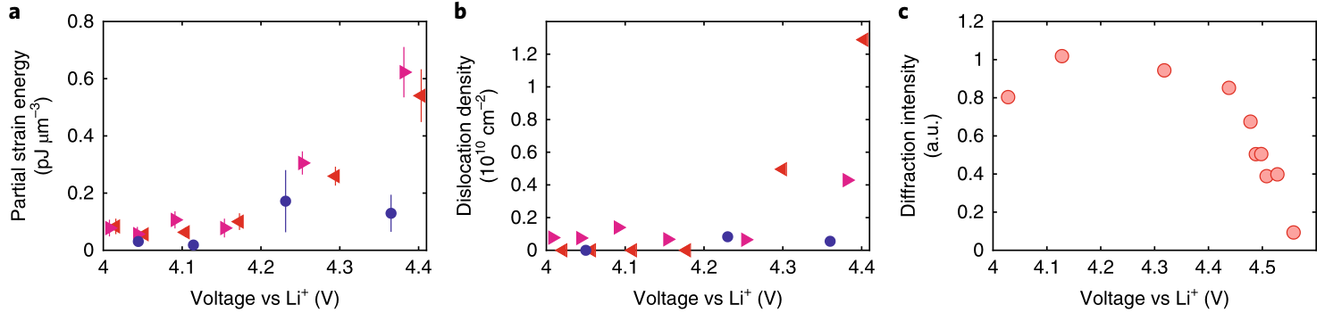
Strain energy landscape of single particles of layered oxides. (a)
Partial strain energy Ep,001 for two LRLO particles
(triangles) and a NCA particle (circles). (c) Operando evolution of the
dislocation density ρ0 for the same particles. (c)
Evolution of the superstructure diffraction peak intensity during the
first charge measured in situ using X-ray diffraction from a large
number of particles. The uncertainties in a represent the standard
deviation between strains calculated using various thresholds for the
reconstructed amplitude in phase retrieval (10 equidistant values
between 0.1 and 0.2).
|
|
Reference:
A. Singer, M. Zhang, S. Hy, D. Cela, C.
Fang, T.A. Wynn, B. Qiu, Y. Xia, Z. Liu, A. Ulvestad, N. Hua, J. Wingert, H.
Liu, M. Sprung, A.V. Zozulya, E. Maxey, R. Harder, Y.S. Meng and O.G. Shpyrko,
"Nucleation of dislocations and their dynamics in layered oxide cathode
materials during battery charging,"
Nature
3, 641-647 (2018). |
Old(er) Research Highlights:
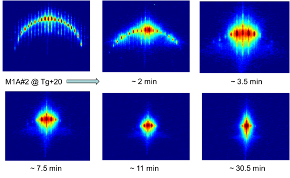
Direct Measurements of Antiferromagnetic Domain Wall Fluctuations
|
Measurements of magnetic noise
emanating from ferromagnets owing to domain motion were first carried out
nearly 100 years ago, and have underpinned much science and technology.
Antiferromagnets, which carry no net external magnetic dipole moment, yet
have a periodic arrangement of the electron spins extending over macroscopic
distances, should also display magnetic noise. However, this must be sampled
at spatial wavelengths of the order of several interatomic spacings, rather than the macroscopic scales
characteristic of ferromagnets.
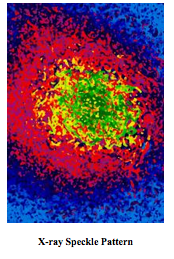
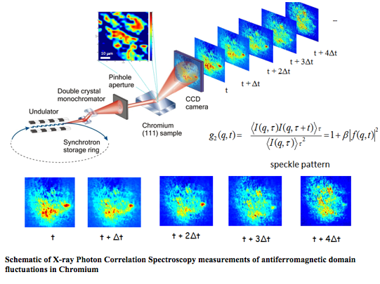
We have used X-ray Photon
Correlation Spectroscopy to couple to the fluctuations in the nanometre-scale superstructure of spin- and charge-density waves associated with antiferromagnetism
in elemental chromium. Coherent X-ray diffraction produces a speckle pattern
that serves as a 'fingerprint' of a particular magnetic domain configuration.
The temporal evolution of the patterns corresponds to domain walls advancing
and retreating over micrometer distances. This work demonstrates a useful
measurement tool for antiferromagnetic domain wall engineering, but also
reveals a fundamental finding about spin dynamics in the simplest antiferromagnet: although the domain wall motion is
thermally activated at temperatures above 100 K, it has a rate that
saturates at a finite value—consistent with quantum
fluctuations—on cooling below 40 K. |
|
|
<
Reference:
O. G. Shpyrko, E. D.
Isaacs, J. M. Logan, Yejun Feng,
G. Aeppli, R. Jaramillo, H. C. Kim, T. F.
Rosenbaum, P. Zschack, M. Sprung, S. Narayanan, and
A. R. Sandy; "Direct measurement of antiferromagnetic domain
fluctuations,"
Nature
447, 68 (2007). |
|
X-ray
Diffractive Imaging of Magnetic Nanoscale Domains
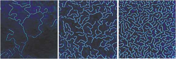
Evolution of magnetic domains in GdFe multilayer films as a
function of applied field.
|
|
We have performed lens-less microscopy of extended magnetic nanostructures, in which a scanned series of dichroic coherent diffraction patterns is recorded and numerically inverted to map its magnetic domain configuration. Unlike holographic methods, it does not require a reference wave or precision optics. In addition, it enables the imaging of samples with arbitrarily large spatial dimensions, at a spatial resolution limited solely by the coherent X-ray flux, wavelength, and stability of the sample with respect to the beam. We demonstrate this approach by imaging ferrimagnetic labyrinthine domains in a Gd/Fe multilayer with perpendicular anisotropy and follow the evolution of the domain structure through part of its magnetization hysteresis loop. In such multilayer systems, alternating layers of a transition metal and a rare-earth metal are deposited to create an artificial ferrimagnet with perpendicular magnetic anisotropy. The balance between exchange, anisotropy, and dipolar energies results in stripe domains. |
|
This type of pattern is commonly found in a
wide class of uniaxially modulated condensed matter
systems, including diblock copolymers, liquid
crystals, Langmuir monolayers, adsorbates at
metallic surfaces, incommensurate structures, membranes, vesicles, and ferrofluids, as well as dewetting,
phase separation, dealloying, and convection
patterns. The
characteristic length scale corresponding to
the width of the stripe is generally defined by the energetic balance
between
short- and long-range interactions
. The ability to tune the relative
strength of these interactions and to control the degree of orientational or translational order in many of these
systems represents a pathway for driven or guided self-assembly at nanoscale.
Reference:
“
Dichroic Coherent
Diffractive Imaging” Ashish Tripathi,
J. Mohanty, S. Dietze,
O.
G. Shpyrko, E. Shipton, E. Fullerton, S.S. Kim and
Ian McNulty, Proc. Natl. Acad. Sci. U S A, 108 (33), 13393-8 (2011)
|
|
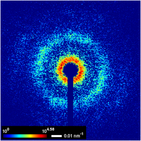
Phase transitions in
Langmuir-Blodgett films
|
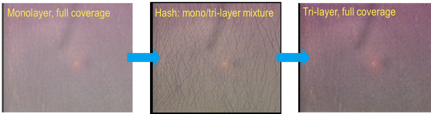
The
optical microscopy images of Au nanoparticle LB film at different phases
|
|
|
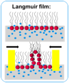
Understanding of the
mechanical and dynamical properties of Langmuir-Blodgett films provides
insights into the dynamics, collapse and relaxation of biological membranes
such as lung surfactant and nanoparticle self-assembled thin films. During
the compression process, the self-assembled nanoparticle Langmuir-Blodgett
films at the air-liquid interface exhibit rich mechanical behavior,
undergoing a rapid structural evolution which is
marked by the phase transition from monolayer to hash to multilayer. We have
performed different synchrotron X-ray scattering techniques to study the
phase transition of these systems. We explored the capability of Grazing
Incidence X-ray Off-Specular (GIXOS) scattering to capture this rapid
structural evolution. The detailed analysis of GIXOS data from the
self-assembled Au nanoparticle films offers the quantitative,
Angstrom-resolution details of electron density profile normal to the surface
with a sub-minute temporal resolution that allows us to study in-situ the
rapid evolution of nanoparticle films structure in response to the lateral
compression. |
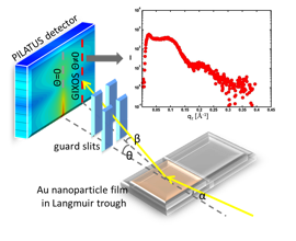
Schematic view of the scattering
geometry with the Langmuir trough. An inset shows a typical intensity
profile
from Au nanoparticle film, plotted as function of wavevector
transfer normal to the surface, qz.
|
|
Morphological instabilities
in Nanoimprinted Polymer nanostructures
The
stability of the imprinted polymer pattern is crucial to the Nanoimprint Lithography technique. The development of a
novel collective type of zig-zag
capillary instability has been recently observed upon annealing of line-space
polymer patterns formed with nanoimprint
lithography. We performed
synchrotron based, time-resolved Grazing Incidence Small Angle X-ray
Scattering (GISAXS) studies of the onset and development of this zig-zag instability that
complement earlier Atomic Force Microscope (AFM) based studies. Time resolved GISAXS provides the
in-situ measurement of the rapid formation of instability during the
annealing process over large sample area, a task that remains challenging for
local scanning probes such as AFM.
A relatively simple unit cell model has been applied to quantitatively
interpret the GISAXS data and identify the onset of the Rayleigh-Taylor
morphological instabilities. |
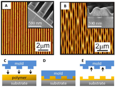
AFM data of as-imprinted polystyrene
with fluorosurfactant line-space grating. SEM (inset) shows the lines
rectangular cross-section at a fractured edge. (B
) After
annealing the sample a
lateral zig-zag instablilty
forms in the pattern. (C-E) Nanoimprint
Lithography schematic.
|
|
Time
evolution of the GISAXS pattern following the evolution of the
Rayleigh-Taylor morphological instabilities
|
|
|
Lateral Diffusion in 2D Nanoparticle
Self-Assembled Films
The
recent trend towards smaller and lighter technological devices has created a
substantial demand for nanoscale films engineered to meet specific optical,
electrical, and magnetic properties.
Examples include the implementation of thin films into antireflective
coatings, computer memory, and lightweight, flexible energy-storage
devices. Despite the number of
potential
applications, which will only continue to grow, methods for characterizing
the mechanical properties and stability of ultrathin films have not been well
established. The drastic increase
in spatial and time resolution achieved by coherent x-ray synchrotron sources
and beamlines in recent years has begun to allow
for direct coupling of x-rays to monitor interparticle
motions.
|
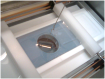
Langmuir-Blodgett
trough setup used for compressing nanoparticle monolayers self-assembled on
the surface of water
|
|
|
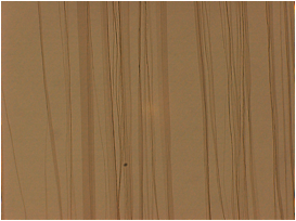
Micron-sized
wrinkles in a compressed 5nm iron oxide nanoparticle monolayer
|
We
study self-assembled monolayers, e.g. iron oxide nanoparticles self-assembled
at the water-air interface using X-Ray Photon Correlation Spectroscopy
(XPCS). This technique
effectively compares “snapshots” of the diffraction pattern produced by the
monolayer at different times in order to reproduce the film structure and interparticle dynamics. These interparticle
motions depend directly upon parameters such as particle size, and evolve as
the surface pressure of the system is increased, ultimately leading to
“buckling” or “wrinkling” at points of local instabilities.
|
|
|
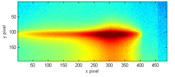
The
first-order Grazing Incidence Diffraction (GID) peak for a self-assembled
monolayer of 20nm iron oxide nanoparticles
|
||
|
Coherent X-ray diffraction imaging of
strained nanowires
Highly
strained nanoscale wires have been intensively studied because a structural
deviation of the crystal from the ideal arrangement can enhance dramatically
electronic, magnetic and optical properties, and its high-surface-to-volume
ratio is useful in a wide range of applications such as
catalysis, sensors, batteries,
fuel cells, and magnetic
devices.
|
||
|
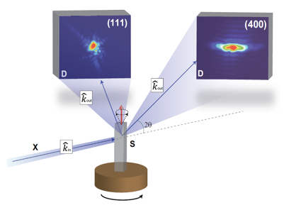
We useCoherentX-ray
Diffraction in the Bragg geometry
highly sensitive to lattice distortion caused by deformation fields within a
small crystal. This technique allows us to image not only the shape in 3D but
also the projection of the crystal lattice distortion on to the Q vector of
the measured Bragg spot.
Upon
successful inversion to real space, the amplitude and phase of the complex
density represents physical density and lattice deformation, as projected
onto the Q vector of the Bragg peak chosen, respectively.
|
|
|
|
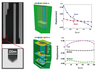
Scanning electron micrograph of the specimen
(left) and
3D reconstruction of the phases and
magnitude
of the displacement
fie
ld of a single crystal
Ni
nanowire
(right)
|
However,
since the detector only measures intensities, a direct reconstruction is
impossible, unless a phase problem is solved via special phasing procedure,
an iterative process using back and forth Fourier transforms between real and
reciprocal space.
Reference:
E.
Fohtung,
J. W. Kim, Keith T.
Chan, Ross Harder, Eric E. Fullerton, and O. G. Shpyrk
o
;
"
Probing the 3D strain
inhomogeneity and equilibrium elastic properties of single
crystal
Ni nanowires"
,
submitted to Applied Physics Letters
(May 2012).
|
|
|
Strain Mediated Magnetoelectric
Coupling as a route to Novel Hybrid Multiferroic
Devices
Smart materials for
sensor technology, non-volatile device memories for information technology,
and ultrasound generators in medical imaging have one thing in common, their
active elements consist of ferroelectrics (FE) driven by voltages or ferromagnetics (FM) driven by magnetization. In the
quest
to design high functionality devices to meet today's consumer technological
demands, high focus has been given to multiferroic.
However, the coexistence of magnetic order and ferroelectric polarization
combined in a single-phase material has proven to be rear as most of these
materials tend to have low magnetic ordering temperatures and are often
antiferromagnets, in which the magnetoelectric (ME)
coupling effect is intrinsically small. F
ield
effect concepts can be applied to complex oxides, such as high-TC
superconductors and manganites, in order to create
new electronic and magnetic phase |
|
|
|
In
magneto-electronic materials such as multiferroics an external electric field
displaces ions from their equilibrium positions, which alters the magnetostatic and exchange interactions yielding magnetoelectric coupling.
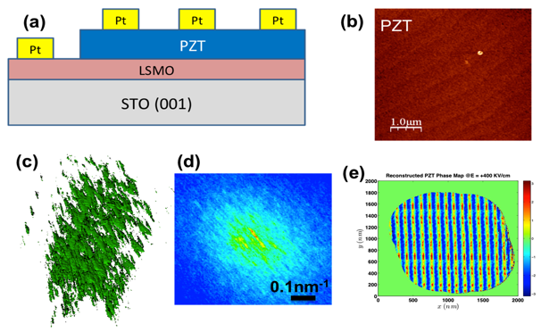
We utilize an
alternative approach to design multiferroic-hybrid
devices (see fig 1a) of FE-FM heterostructures where the magnetoelectric
coupling emerges from strain-mediated interaction between individual phases.
Specific examples of systems we are presently studying and intend to pursue
further include but not limited to heterostructures consisting of magnetic (LSMO, transition-metal alloy)
filmsand
highly piezoelectric (PMN-PT and PZT) layers. The devices schematic structures are
shown in Fig. 1a. The sample
structure is Si (001) substrates, STO (SrTiO3), LSMO
(La1-x
Sr xMnO3) bottom
electrodes, PMN-PT((1-x)[Pb(Mg1/3Nb2/3)O3]-x[PbTiO3]
or PZT (PbZrTi3) and a top electrode that is either a non-magnetic
metal (e.g. Pt)
or a magnetic transition metal film (e.g.
Co). In the presence of an external applied electric field, Ti4+(
Zr
4
+
)
cations
are formed as the Ti- atoms are displaced from their equilibrium
position. This alters the
magnetostatic
and exchange interactions yielding magnetoelectric
coupling. Strain is observed as displacement derivatives.
This strain mediation
is qualitatively studied in the form of a nonlinear thermodynamic theory for
strain-mediated direct Magnetoelectric effect. We
utilize
our in-house atomic force microscopy (AFM) and Piezoelectric Force Microscopy
(PFM) to understand the switching nature of the Ferroelectric domains. To
understand how it couples to the lattices and its depth dependence, Bragg
Ptychographic Coherent Diffraction Imaging (BCDI) is invoked. The interfacial
coupling and enhancement of magnetization is probed using neutron scattering
techniques.
Our complementary use
of various probing techniques enables us to nondestructively obtain insights
to the coupling phenomena of the order parameters required for device
functionality. We use Bragg Ptychographic Coherent Diffraction Imaging (BCDI)
and neutron scattering as the unique tools of choice for sub-nanometer
resolution and nondestructive probing of the order parameters in these
materials and devices.
|
|

|

|

|
Our research summarized in a song ("I Study Chromium") by REU student Ernesto Abruna:
-
My LSXS Lecture Notes
have been posted here:
